
During profound sleep, breathing and brain signals become disorganised.
Breathing signals are disrupted by deep sleep.

Researchers have shown that during the deepest stage of sleep, respiration and brain activity in important movement circuits become out of sync. This division changes how the sleeping brain interprets signals from the body and uncovers a secret deep rest rule.
Breathing signals are disrupted by deep sleep.
During the deepest sleep, breathing no longer determines the timing of electrical cycles recorded from movement circuits deep within the brain. Dr. Bon-Mi Gu of Hackensack Meridian Health (HMH) analysed those signals and showed that as sleep deepens, the breathing-brain connection inside motor networks drastically diminishes.
During awake and light sleep, the same circuits closely monitor breathing; but, as the brain enters the slowest sleep patterns, this coordination diminishes. The way that deep sleep alters communication between internal circuits and the body may be explained by knowing why the brain releases that timed signal.
Brain messages are guided by breathing rhythm.
Neural impulses can be timed by breathing, and a 2017 review traced this connection from deep brain networks to the nose. This breath-linked time between breathing and brain activity is referred to by researchers as respiration-neural coupling, and it frequently intensifies during alert states.
Coupling did not act consistently throughout Dr. Gu's data, suggesting that the brain uses local rules. Monitoring sleep and anaesthesia could be more accurate if we know where that timing holds and where it drops off.
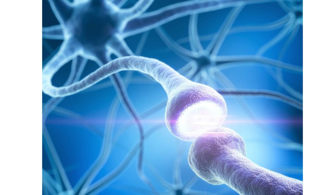
Circuits that move the brain
Signals originated from the motor cortex and the substantia nigra, a deep brain area related to movement control. The substantia nigra is located inside the basal ganglia, which are deep brain hubs that aid in initiating and stopping movements, according to neuroscientists.
The team was able to investigate whether breathing timing diffused across both layers of movement control by pairing these deep signals with the motor cortex. Numerous brain regions are affected by breathing rhythms, therefore alterations in these motor hubs may have an impact on movement and sleep issues.
Comparing different stages of sleep
Through peaceful wakefulness, various sleep stages, and anaesthesia, the team observed brain signals and diaphragm activity for each mouse. The brain exhibits slow waves and less movement during non-REM sleep, the tranquil stage that encompasses deep sleep.
Later, twitchy muscles and the type of brain activity associated with dreams are brought on by REM sleep, which is characterised by fast eye movements. The researchers was able to distinguish between changes related to anaesthesia medicines and changes related to sleep itself by tracking the same mouse under several settings.
Sleep that impairs timing
The weakest breath-to-brain time was caused by non-REM sleep, and both recorded regions demonstrated the decline. Breathing was less frequently associated with electrical activity in the substantia nigra and motor cortex as compared to silent wakefulness and REM sleep.
Instead of only changing pace with each deeper stage, the coupling weakened as REM sleep gave way to non-REM sleep. The pattern suggests that the deepest rest state alters the way the brain responds to bodily rhythms.
The narrative is altered by anaesthesia
Even when the animals appeared still, their respiration and brain activity did not resemble sleep under anaesthesia. The substantia nigra demonstrated significantly stronger coupling with anaesthetic medications that reduce brain activity and reflexes.
Signals from the motor cortex did not increase in strength.
This discrepancy suggests that one circuit may be overdriven by anaesthesia while another circuit remains more in line with typical sleep processing.
Interaction between motor circuits
Stronger synchronisation between the substantia nigra and the motor cortex accompanied slow delta waves. Breathing had fewer opportunities to synchronise their electrical signals when those two regions aligned with one another.
This pattern implies that, even if it means losing contact with the body, deep sleep promotes internal communication across motor circuits. Scientists could target that physiological link to modify sleep depth without altering breathing if more research verifies the cause.
Prospects for future research
Parts of the basal ganglia are damaged by Parkinson's disease, and many patients experience breathing difficulties at night in addition to disturbed sleep. Changes in breath-brain timing within that system could be used to monitor early stress on those circuits because the substantia nigra is located there.
In order to ensure patient safety, clinical teams now monitor breathing as they sleep, and new research indicates that brain rhythms may provide an additional layer. Although mouse data alone cannot predict human symptoms, it provides a clear circuit that can be tested in subsequent patient research.
The study reframes a fundamental body-brain handshake by demonstrating how deep sleep can sever the time connection between breathing and important motor circuits.
It will be necessary to confirm when coupling is beneficial, when it is detrimental, and how medications affect it in humans and other brain regions.
About the Creator
Keep reading
More stories from Francis Dami and writers in Humans and other communities.
After two millennia, the Commodus passage is once again accessible to the general public.
Visitors to Rome are exploring the Commodus Passage, a secret passageway in the Colosseum that was formerly only accessible to emperors, for the first time in nearly two millennia.
By Francis Damiabout a month ago in Humans
Ways To Get Over A Breakup Fast Using Emotional Strategies
The first process to the healing phase of a breakup is the ability to be self-aware about how you feel. The inhibition of sadness, anger or frustration increases emotional suffering. It is necessary to allow these emotions so that you could feel them and process them to heal.
By Willian James5 days ago in Humans


Comments
There are no comments for this story
Be the first to respond and start the conversation.